Indian Journal of Physiology and Pharmacology
Volume 63 - Number 1 January - 2019

Medical Education / Method Paper

Demonstration of Nerve Muscle Preparation in Rats: For Nerve Muscle Physiology eaching

Arpita Chakraborty, Ashish Arvind, Shival Srivastav, Geetanjali Bade, Simran Kaur and Suman Jain*

Department of Physiology, All India Instittue of Medical Sciences, Ansari Nagar, New Delhi, India

*Corresponding author :
Prof. Suman Jain, Department of Physiology, AIIMS, New Delhi,
Email: sumanjain10@gmail.com

Abstract

Purpose of the study: Physiology practical complement learning of concepts taught in didactic theory lectures. Therefore, they are instrumental to comprehensive learning. Learning of physiology of excitable tissues is supplemented by observing the same phenomena in action in the laboratory. We developed an in-situ rat nerve-muscle model to demonstrate nerve-muscle physiology phenomenon to undergraduate and postgraduate students. This mimics the physiological conditions unlike the Rana tigrina experiments which were done earlier. Further, frequent unavailability of frogs envisaged us to find an alternative for demonstration of these concepts.

Materials and methods: We developed and standardized an in-situ rat soleus muscle-sciatic nerve model to demonstrate the phenomenon of nerve-muscle properties like simple muscle twitch, effect of strength of stimulus, effect of two successive stimuli and tetanus to the undergraduate and postgraduate students at our institute.

Results: Simple muscle twitch was recorded and effect of increasing strength of stimuli was observed. Thereafter, effect of two successive stimuli and genesis of tetanus was recorded in the same experimental set up. We successfully standardised this in-situ model for teaching nerve and muscle physiology to our students.

Conclusion: We propose an easy and effective experimental rat in-situ model for nerve-muscle physiology teaching to students.


Introduction

Basic sciences such as physiology serve as the foundation upon which a medical student builds his understanding of medical science. The knowledge of physiology serves as a cornerstone for understanding pathophysiology and treatment strategies.

Didactic lectures and practical sessions constitute two basic tenets of physiology teaching. Newer modalities such as group based learning, problem based learning and role-playing has emerged over the years to garner student attention and facilitate engagement in the learning process. While such modalities serve to provide theoretical concepts, the age old dictum of “I hear and I forget, I see and I remember, I do and I understand” still hold true.

To achieve this end, practicals were included in the medical curriculum to strengthen students’ understanding of concepts by individual and demonstration practical exercises. Animal models have been employed for demonstration of various concepts of nerve and muscle physiology. Sciatic nerve-gastrocnemius muscle model of Rana tigrina was a standard and effective learning tool for strengthening of nerve muscle physiology concepts. Since the availability of the above model has declined due to multiple reasons, the practical teaching in this regard has receded to explanation of graphs using chalk and talk or power- point methods which fail to capture the students’ interest.

Therefore, there is an urgent need to establish another animal model for nerve-muscle physiology practical exercise that is easily available, ethically approved and closely mimics physiological properties of human neurophysiology to serve as an effective teaching aid.

Since rat is a mammal, it has similar physiological principles governing its’ body function as humans. The rats are easily available and easy to breed. They are also relatively harmless and easy to handle for performing the laboratory practical for undergraduate students. Moreover they are commonly used as animal models for neurophysiology and metabolic studies. So, they have potential to be used for elucidating nerve muscle physiology concepts for medical teaching.

We addressed this lacuna by developing an in-situ soleus muscle - sciatic nerve model in rat for demonstration of practical physiology concepts.

Materials and Methods

Materials required

The equipment required for this practical exercise included Wistar rat, Digital Data acquisition system, stimulating hook electrodes, force transducer, dissecting instruments, dissection board, weights, saline solution, anaesthetic agent.

Objectives addressed

We addressed the following learning objectives in our study:

  1. Determination of subthreshold, threshold, suprathreshold, maximal and supramaximal strength of stimuli.
  2. Recording simple muscle twitch by stimulating the motor nerve using a supramaximal stimulus
  3. Determination of the effects of two successive stimuli and studying how inter-stimulus interval may affect muscle contraction
  4. Demonstration of the phenomenon of incomplete and complete tetanus

Adult male Wistar rats (body weight 200-250 gm) were obtained from institutional (All India Institute of Medical Sciences, New Delhi, India) central animal facility. The protocol was approved by Institute’s Animal Ethics Committee (65/IAEC-1/2018). Powerlab 4T™ system (AD Instruments, Australia) was used for data acquisition. Data was displayed in real time using LabChart™ software version 8.1 (AD Instruments, Australia) installed on a desktop system. Data was stored for offline analysis later. Stimulating hook electrodes were used to deliver electrical stimuli to the nerve. The detailed procedure is described as follows:

Calibration:

Since the transducer measures contraction strength in Volts by default, it is essential to calibrate the same before the start of the experiment to get values in grams. Transducer channel was identified and zeroing was done before commencement of calibration. Two-point calibration was done by applying weights from 10-50 g in a step up and step down manner. Unit conversion was done to get the subsequent values in Newton.

Animal preparation:

After setting up the data acquisition system, animal dissection was performed. Adult male Wistar rats weighing approximately 200-250 grams were procured from the Institute Animal Housing facility. They were housed in departmental animal house in a temperature-controlled room at 24±2%C with a light: dark cycle of 14:10 hours and provided ad libitum food and water. Six hours of fasting was done before the start of dissection. It was weighed and glycopyrrolate injection (0.5 mg/kg IM) was given to prevent secretions. After 5-10 min, thiopentone at a dose of 50 mg/kg was injected intraperitoneally and it was covered to keep away from direct light. Once the rat was anaesthetized, it was placed on dissection board and its limbs were tied to mounting nails with the help of thick cotton thread to prevent any movement taking care not to injure the skin. Depth of anesthesia was checked by eliciting withdrawal reflex.

Animal dissection:

In anaesthetized rats incision was given on the dorsal side of right thigh extending up to ankle joint. By careful blunt dissection, gastrocnemius muscle was exposed and subsequently soleus was separated from its surrounding attachments which lie below it. Soleus muscle is flat and red in appearance and lies along the surface of tibia. After the identification of soleus muscle, it should be detached from gastrocnemius and plantaris muscle by dental scalpel. As the tendon of three above mentioned muscles are attached, tendon of soleus muscle should be isolated from them without damaging any blood vessels. Origin and insertion of soleus were kept intact. A cotton thread was tied to the tendon of soleus muscle and other end was attached to the force transducer. Extending the incision proximally up to the vertebral column, sciatic nerve and its branches were exposed and separated from surrounding connective tissue with the help of glass seeker. Hook stimulating electrodes were applied to the sciatic nerve. Liquid paraffin was applied to exposed nerve to insulate and prevent evaporative loss. Entire area was covered with gauge piece moistened with mammalian ringer. Fig. 1A and 1B shows the representative graphs of the set up for the experimental protocol.

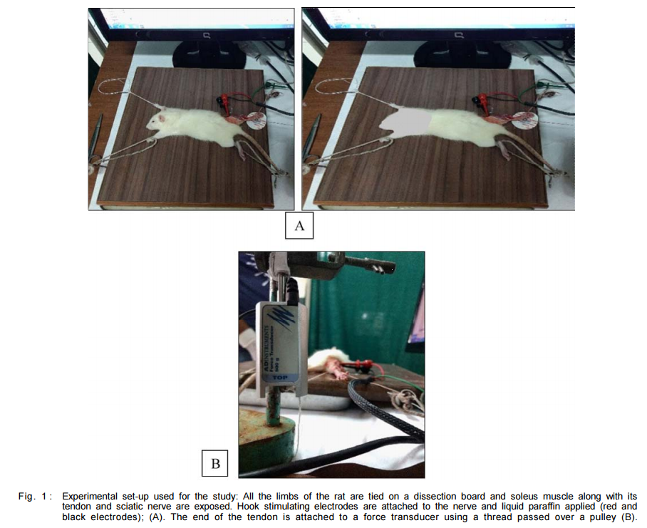
Stimulation and data acquisition:

The tendon of the soleus muscle was isolated and tied to a force transducer with the help of a cotton thread. Sciatic nerve was placed on the stimulating hook electrodes as shown in Fig. 1B. Square wave pulse of 1 ms duration with varying strength was delivered.

Recording of threshold stimulus intensity of simple muscle twitch (SMT):

To determine the threshold, square wave pulses of fixed duration and increasing current intensities was used to stimulate the sciatic nerve. Current intensity was gradually increased from 0.1V in increments of 0.1V till a twitch was elicited. Minimal stimulus intensity which elicited the simple muscle twitch was considered as threshold intensity of SMT. The peak tension generated during a simple muscle twitch was calculated. The latent period, contraction and relaxation periods were also analyzed.

Recording of suprathreshold, maximal and supramaximal stimulus:

Current intensity beyond the threshold intensity was gradually increased till a twitch with maximum amplitude was obtained. Further increase in the current intensity does not increase maximum amplitude of simple muscle twitch. This value was considered as suprathreshold intensity of SMT and stimulus is supramaximal. Peak tension generated at the time of contraction was calculated. The latent period, contraction and relaxation period were analyzed.

Recording of effect of two or more successive stimuli:

If a skeletal muscle is given two successive stimuli of supra-threshold intensity, the response to the second stimulus depends upon the time interval between the two stimuli. Inter-stimulus intervals are set by altering the frequencies. In same nerve-muscle preparation when two supramaximal stimuli are applied in such a manner that the second stimulus falls in the following phases:

  1. Completion of first twitch
  2. End of relaxation of the first twitch
  3. During relaxation of the first twitch
  4. During contraction period of the first twitch
  5. During second half of the latent period
  6. During first half of the latent period

Inter-stimulus intervals or frequency of stimulation were calculated by analyzing latent period, contraction period and relaxation period of simple muscle twitch. At each of the two successive stimuli, twitch amplitude and duration were recorded. The shape of the SMT curve was also analyzed during different phases as described above.

Genesis of Tetanus:

If a skeletal muscle is repeatedly stimulated at such a frequency, so that, relaxation period of one twitch is superimposed with its previous one and muscle does not get relaxed, a state of sustained contraction is obtained which is known as “complete tetanus”. Below that frequency muscle relaxation occurs incompletely and it is considered as “incomplete tetanus”. For the genesis of tetanus, sciatic nerve is stimulated at supra-maximal stimulus intensity with increasing frequency. Amplitude of contraction and peak tension generated at different frequencies were calculated. Force-frequency relationship of muscle was plotted and analyzed.

Results

In the null-model, ICC was found to be 0.24, which is the proportion of variance in the outcome score that is attributable to examiner level factors. It can be assumed here that part of this variance may be due to the different methods of assessment. For that, we also calculated the ICC from the full-model, which came out to be 0.01. Thus, the decrease in variance due to examiners between the models can be accounted to different methods of assessment.

We also calculated the R - squared from the variance component of both the models and it was found that 96% of the variance was explained by use of different methods of assessment. P-value was also statistically significant (<0.05) for the fixed effect of the methods of assessment.

Pearson’s correlation coefficient between the two semi-structured assessment was 0.754 as compared between conventional and semi-structured assessment which was found to be 0.488 and 0.466.


Results

The representative graph records are depicted in Fig. 2 to 6. Simple muscle twitch was recorded and effect of increasing strength of stimuli was observed. Effect of two successive stimuli and genesis of tetanus was recorded in continuation of the same set of experiment. Force-frequency graph was plotted from the obtained data.

Discussion

The present paper proposes an innovative and effective method for teaching nerve muscle physiology to undergraduate and postgraduate students. It also provides a substitute to frog experiments and simulation techniques. Handling and dissecting the rat nerve-muscle preparation imparts a set of surgical skills and handling of tissue and keeping them viable through experiment helps to develop team work and cooperation. Effect of various physical, physiological and pharmacological conditions can also be studied in the same set of experiments.

Assessment of students can be done on individual performance basis for postgraduates and as OSPE based on graphs and different set of physiological conditions for undergraduates.

Limitation of these experiments is few if proper protocol and precautions are followed. Long term anaesthesia leads to fall in body temperature of rat which has been leading cause of death of rats during experiments. Maintaining body temperature by using heating pad or other devices can help to overcome this limitation.

Further scope for this experimental set up is to extend it to develop models to mimic the effect of exercise, training and nerve muscle injury.

References

  1. MacIntosh BR, Esau SP, Holash RJ, Fletcher JR. Procedures for Rat in situ Skeletal Muscle Contractile Properties. JoVE J Vis Exp 2011; 56: e3167– e3167.
  2. Burke RE, Levine DN, Tsairis P, Zajac FE. Physiological types and histochemical profiles in motor units of the cat gastrocnemius. J Physiol 1973; 234(3): 723–48.
  3. Norenberg KM, Fitts RH. Contractile responses of the rat gastrocnemius and soleus muscles to isotonic resistance exercise. J Appl Physiol 2004; 97(6): 2322–2332.9.